
Which of the Following is NOT a Motor Cranial Nerve? Understanding Cranial Nerve Functions
Table of Contents
- Introduction: The Cranial Nerves at a Glance
- Quick Answer: The Non-Motor Cranial Nerves
- The Purely Sensory Cranial Nerves (Non-Motor)
- CN I: Olfactory Nerve (Smell)
- CN II: Optic Nerve (Vision)
- CN VIII: Vestibulocochlear Nerve (Hearing and Balance)
- The Purely Motor Cranial Nerves (For Contrast)
- CN III: Oculomotor
- CN IV: Trochlear
- CN VI: Abducens
- CN XI: Accessory
- CN XII: Hypoglossal
- The Mixed Cranial Nerves (Both Sensory and Motor)
- CN V: Trigeminal
- CN VII: Facial
- CN IX: Glossopharyngeal
- CN X: Vagus
- How I Remember Them: Simple Mnemonics That Stick
- Summary Table: All 12 Cranial Nerves and Function Types
- Clinical Relevance: Why This Classification Matters in Real Life
- How I Test Cranial Nerves in a Quick Neuro Exam
- Common Confusions and Exam Traps I See Often
- A Simple Engineering Analogy That Helped Me
- Key Takeaways
Introduction: The Cranial Nerves at a Glance
I remember the first time I sat with a skull model and a set of flashcards. Twelve pairs of cranial nerves stared back at me. Some moved eyes. Others made faces smile or helped you swallow. A few did nothing but carry sensory information from your world into your brain. Once I grouped them by function everything clicked.
We classify cranial nerves by what they do:
- Motor: move muscles and control glands
- Sensory: carry sensation like smell, vision, hearing, balance, taste, or touch
- Mixed: do both motor and sensory
You do not have to memorize the whole brainstem in one sitting. You only need a clean mental map that separates sensory vs motor vs mixed. Then you build the details on top.
Quick Answer: The Non-Motor Cranial Nerves
If your question is “Which of the following is not a motor cranial nerve?” here is the straight answer.
- Olfactory nerve (CN I) — sensory only
- Optic nerve (CN II) — sensory only
- Vestibulocochlear nerve (CN VIII) — sensory only
These three carry special sensory input. They do not move muscles. They do not drive glands. They are not motor.
The Purely Sensory Cranial Nerves (Non-Motor)
Here is why CN I, II, and VIII qualify as non-motor. I will share what I look for in real cases so the details stick.
CN I: Olfactory Nerve (Smell)
What it does: CN I carries the sense of smell from olfactory receptors in your nose to your olfactory bulb then on to higher brain regions. That pathway lets scents trigger memories and emotions fast.
Why it is not motor: It provides special sensory input only. No muscle fibers. No motor output.
In practice: When I see a patient who lost their sense of smell after a viral illness I think of CN I. Anosmia is common and it ranges from mild to severe. Many people lost smell temporarily after COVID-19 and a large portion recovered over weeks to months. I always check that it is not due to nasal blockage first.
Tip to remember: “Olfactory = odor.” Smell only.
CN II: Optic Nerve (Vision)
What it does: CN II carries visual information from the retina to the brain. It sends signals about light, color, shape, and movement.
Why it is not motor: It is a sensory highway. No muscles attach to it and no motor fibers run within it.
Clinical pearls I use:
- Field cuts suggest where along the visual pathway the lesion sits
- Optic neuritis can show up with painful eye movement and vision loss
- About half of people with multiple sclerosis have optic neuritis at some point
- Glaucoma damages the optic nerve slowly and it is a leading cause of irreversible blindness
Common exam trap: The pupillary light reflex involves CN II and CN III. CN II senses the light. CN III constricts the pupil. Students sometimes call CN II “motor” because the pupil constricts in that reflex which is understandable. The motor part belongs to CN III. CN II stays sensory.
CN VIII: Vestibulocochlear Nerve (Hearing and Balance)
What it does: CN VIII carries sound from the cochlea and balance signals from the vestibular apparatus. That includes the semicircular canals and otolith organs. It helps you stand upright and move your eyes with your head so your world stays steady.
Why it is not motor: It is pure special sensory. No muscle groups receive commands from this nerve.
What I see in clinic:
- Sensorineural hearing loss grows more common with age. A large chunk of people over 65 notice it
- Vertigo, tinnitus, and disequilibrium often trace back to vestibular causes
- Vestibular neuritis presents with sudden severe vertigo and imbalance. The room spins and nausea hits hard
Tip to remember: “VIII” looks like two stacked circles to me which I linked to ears and balance. Silly maybe but it stuck.
The Purely Motor Cranial Nerves (For Contrast)
These nerves move things. They drive muscles or control parasympathetic output. When I think of motor nerves I picture movement and reflexes not senses.
CN III: Oculomotor
What it does:
- Moves most extraocular muscles
- Lifts the eyelid (levator palpebrae)
- Constricts the pupil and accommodates the lens via parasympathetic fibers
Clinical clues:
- A CN III palsy causes a “down and out” eye with ptosis and often a dilated pupil
- I watch the pupil closely. A blown pupil suggests compressive lesions like aneurysm
CN IV: Trochlear
What it does:
- Innervates the superior oblique muscle
- Helps you look down and in
What patients say:
- Vertical diplopia
- Trouble walking downstairs
- They tilt their head to compensate
CN VI: Abducens
What it does:
- Innervates the lateral rectus
- Abducts the eye
Why I never forget CN VI:
- It has a long intracranial course so it is vulnerable to elevated intracranial pressure
- A palsy gives horizontal diplopia and medial deviation of the eye
CN XI: Accessory
What it does:
- Motor to sternocleidomastoid and trapezius
- Turns the head and shrugs the shoulder
Practical tip:
- I test head turn against resistance and shoulder shrug
- Injury sometimes follows neck surgery and shoulder droop stands out
CN XII: Hypoglossal
What it does:
- Motor to tongue muscles
What I look for:
- Tongue deviation toward the side of a lesion on protrusion
- Fasciculations and atrophy in lower motor neuron lesions
- Dysarthria and trouble handling food
The Mixed Cranial Nerves (Both Sensory and Motor)
Four cranial nerves carry both sensory and motor fibers. I group them by what I can test fast.
CN V: Trigeminal
Sensory:
- Touch, pain, and temperature from the face, cornea, nasal mucosa, and oral cavity
- Sensory to anterior two thirds of the tongue (general sensation not taste)
Motor:
- Muscles of mastication: masseter, temporalis, medial and lateral pterygoids
- Tensor tympani, tensor veli palatini, mylohyoid, anterior belly of digastric
Clinical features I watch:
- Trigeminal neuralgia causes severe lancinating facial pain that comes in bursts
- Loss of corneal reflex if V1 sensory input is gone
- Weakness in jaw clench points toward motor branch issues
CN VII: Facial
Sensory:
- Taste from anterior two thirds of the tongue
- A tiny bit of external ear sensation
Motor:
- Muscles of facial expression
- Parasympathetic to lacrimal glands and salivary glands (submandibular and sublingual)
- Stapedius muscle in the ear
At the bedside:
- Bell’s palsy causes acute unilateral facial paralysis
- I look for loss of forehead wrinkling which separates peripheral from central lesions
- Hyperacusis can show up due to stapedius involvement
- Dry eye or mouth points to parasympathetic fibers
CN IX: Glossopharyngeal
Sensory:
- Taste from posterior one third of the tongue
- Sensation from pharynx and posterior tongue
- Carotid body and sinus chemo and baroreceptors
Motor:
- Stylopharyngeus muscle for swallowing
- Parasympathetic fibers to the parotid gland
Clinically:
- Loss of gag reflex can involve IX afferent limb
- Pain can radiate to the ear in glossopharyngeal neuralgia
- Often affected with CN X in bulbar palsies
CN X: Vagus
Sensory:
- Sensation from pharynx and larynx
- Visceral sensory from thoracic and abdominal organs
- Some taste around the epiglottis
Motor:
- Pharyngeal and laryngeal muscles for swallowing and voice
- Parasympathetic output to heart, lungs, and gut
Red flags I listen for:
- Hoarseness suggests recurrent laryngeal nerve involvement
- Dysphagia and nasal regurgitation point toward palatal weakness
- Autonomic symptoms like heart rate irregularities or GI motility issues may appear
How I Remember Them: Simple Mnemonics That Stick
You want mnemonics that work under pressure. These two have saved me more than once.
- For names in order (CN I to XII): “Oh Oh Oh To Touch And Feel Very Good Velvet Ah Heaven.” It sounds goofy but it works
- For function type (Sensory, Motor, Both): “Some Say Marry Money But My Brother Says Big Brains Matter More.” Map each word to CN I through XII in order. S = sensory. M = motor. B = both
Here is how that mapping lands:
- CN I S
- CN II S
- CN III M
- CN IV M
- CN V B
- CN VI M
- CN VII B
- CN VIII S
- CN IX B
- CN X B
- CN XI M
- CN XII M
You will notice CN I, II, and VIII are the sensory outliers. That is exactly the set that is not motor.
Summary Table: All 12 Cranial Nerves and Function Types
Below is the quick-reference table I wish I had during my first neuro exam practice session.
| Number | Name | Function Type | Core Modalities | Quick Clinical Notes |
|---|---|---|---|---|
| CN I | Olfactory | Sensory | Special sensory (smell) | Anosmia after viral infections is common. Check for obstruction first |
| CN II | Optic | Sensory | Special sensory (vision) | Visual field defects map the pathway. Optic neuritis common in MS |
| CN III | Oculomotor | Motor | Somatic motor to eye muscles, parasympathetic to pupil/lens | “Down and out” eye, ptosis, dilated pupil in palsy |
| CN IV | Trochlear | Motor | Somatic motor to superior oblique | Vertical diplopia. Trouble reading or going downstairs |
| CN V | Trigeminal | Both | General sensory face, somatic motor mastication | Trigeminal neuralgia causes severe facial pain |
| CN VI | Abducens | Motor | Somatic motor to lateral rectus | Medial strabismus and horizontal diplopia |
| CN VII | Facial | Both | Taste anterior 2/3 tongue, facial expression, parasympathetic glands | Bell’s palsy, hyperacusis, dry eye/mouth |
| CN VIII | Vestibulocochlear | Sensory | Special sensory (hearing, balance) | Hearing loss, tinnitus, vertigo, disequilibrium |
| CN IX | Glossopharyngeal | Both | Taste posterior 1/3 tongue, pharynx sensation, swallowing, parotid | Dysphagia, loss of gag reflex, ear pain |
| CN X | Vagus | Both | Visceral sensory, pharynx/larynx, parasympathetic to thorax/abdomen | Hoarseness, dysphagia, autonomic issues |
| CN XI | Accessory | Motor | SCM and trapezius | Weak head turn and shoulder shrug after neck surgery |
| CN XII | Hypoglossal | Motor | Tongue movement | Tongue deviates toward lesion. Fasciculations in LMN lesions |
Clinical Relevance: Why This Classification Matters in Real Life
I never learn anatomy in a vacuum. It always ties back to patients and problems.
- Smell and taste complaints spiked after pandemic waves. Many people lost smell or taste temporarily. CN I and the central processing of olfaction sat at the heart of that story
- Vision loss can be subtle or dramatic. Field testing and fundus exams point to CN II and beyond. Optic neuritis, glaucoma, and ischemic optic neuropathy live here
- Diplopia has a pattern. CN III, IV, and VI palsies create distinct eye movement deficits. When someone says the world doubles as they look sideways I think CN VI. When words jump line to line I think CN IV
- Facial paralysis tells a story. CN VII lesions show up on one side with forehead involvement in peripheral palsy. Central lesions spare the forehead because of bilateral cortical input
- Swallowing issues and hoarseness scream CN IX and X. I listen to the voice. I watch the palate elevate. I test the gag reflex gently
- Shoulder droop after neck surgery points to CN XI. Weights feel harder to lift and the scapula wings a bit
- Slurred speech and trouble moving the tongue point to CN XII. I have the person stick out their tongue and I watch which way it points
These distinctions guide testing. They also shape referrals and imaging decisions. If I suspect an aneurysm compressing CN III I act fast. If I see isolated anosmia without red flags I manage expectantly and set expectations.
How I Test Cranial Nerves in a Quick Neuro Exam
You can run through cranial nerves in minutes once you get a rhythm. Here is my approach.
- CN I (Olfactory): I ask about smell change. If needed I use non-irritant scents like coffee or vanilla. I avoid alcohol swabs which stimulate trigeminal nerve endings
- CN II (Optic): I check visual acuity with a near card. I test visual fields by confrontation. I look at the pupils and the optic disc if I have an ophthalmoscope
- CN II and III (Pupils): I shine a light to check direct and consensual responses. CN II senses light. CN III drives constriction
- CN III, IV, VI (Eye movements): I draw an H in the air and watch the eyes follow. I look for nystagmus, diplopia, and any gaze palsy
- CN V (Trigeminal): I test light touch in V1, V2, and V3 distributions. I tap the jaw jerk if upper motor neuron signs are suspected. I test the corneal reflex with a wisp if indicated
- CN VII (Facial): I ask for a big smile, tight eye closure, and eyebrow raise. I watch for asymmetry and forehead involvement
- CN VIII (Vestibulocochlear): I ask about hearing and balance. I use finger rub or whispered voice. I consider tuning fork tests if I need to separate sensorineural vs conductive loss
- CN IX and X (Glossopharyngeal and Vagus): I listen to the voice. I ask them to say “ah” and watch for symmetric palate elevation. I test the gag reflex if needed
- CN XI (Accessory): I test head turn and shoulder shrug against resistance
- CN XII (Hypoglossal): I ask them to stick out the tongue and move it side to side. I check for deviation and fasciculations
Reflex Pathways I Keep Straight
- Pupil light reflex: Afferent CN II, efferent CN III
- Corneal reflex: Afferent CN V1, efferent CN VII
- Gag reflex: Afferent CN IX, efferent CN X
- Jaw jerk: A brisk jaw jerk points toward an upper motor neuron lesion involving trigeminal pathways
- Stapedius reflex: CN VII stabilizes the stapes. Injury can cause hyperacusis
Common Confusions and Exam Traps I See Often
I made every one of these mistakes early on. You do not have to.
- Calling CN II “motor” because of the pupillary response. CN II senses light yet CN III moves the iris sphincter
- Forgetting that CN V provides general sensory to the anterior two thirds of the tongue yet taste there belongs to CN VII
- Missing CN IX taste and sensation in the posterior one third of the tongue
- Confusing mixed nerves with motor only. CN V, VII, IX, and X all carry sensory and motor fibers with parasympathetic components in VII, IX, and X
- Overlooking the parasympathetic role of CN III, VII, IX, and X. Parasympathetics are motor output to glands and smooth muscle even though they feel different from skeletal muscle actions
- Not checking for forehead sparing in facial weakness. It helps separate central from peripheral facial nerve lesions fast
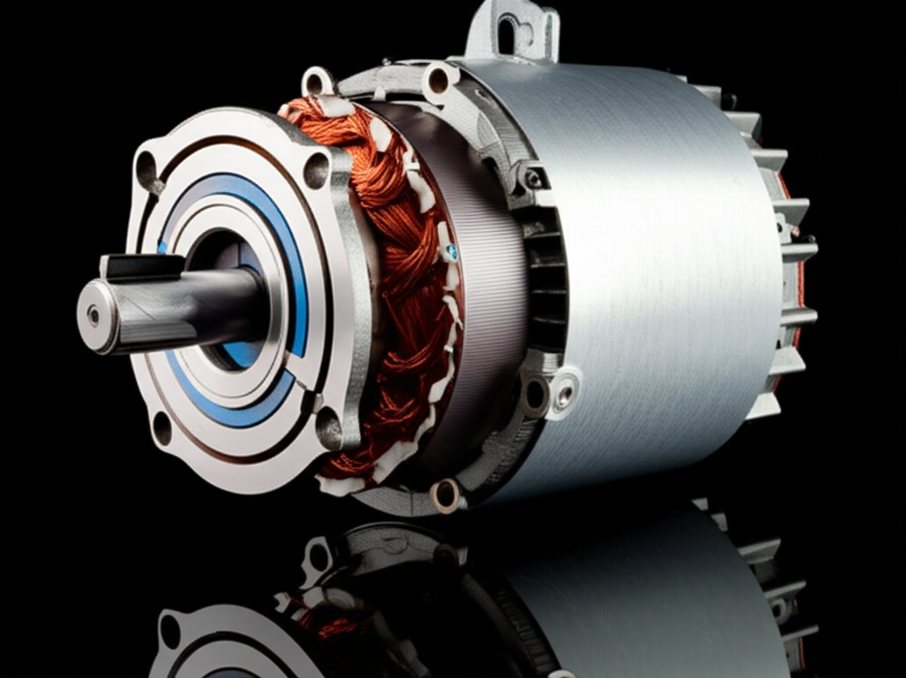
A Simple Engineering Analogy That Helped Me
When I struggled to keep “sensory vs motor” straight I thought about machines. Sensors detect. Motors move. The brain and body do the same dance.
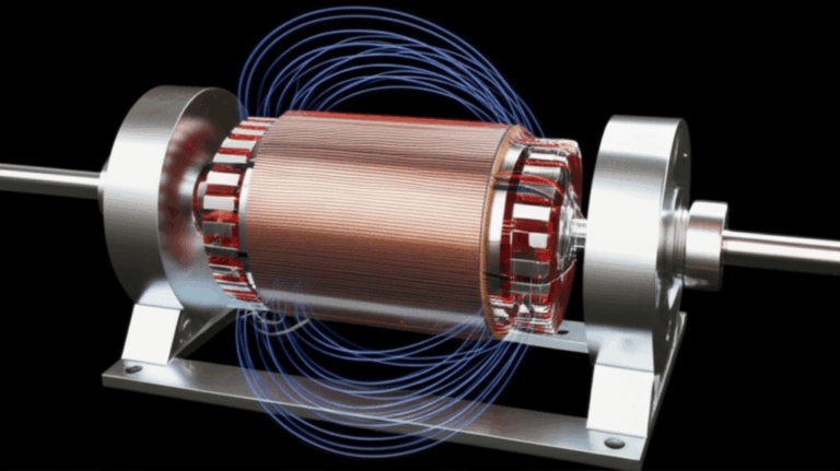
- If you want a quick refresher on what a motor does mechanically you can skim this short explainer on the motor principle. It helped me anchor the word “motor” to motion and output
- Inside many machines you will find a stator and rotor. One stays put. The other spins. I pictured eye muscles like rotors controlled by motor cranial nerves and that image stuck
- Quality matters in machines and bodies. Just like the integrity of motor core laminations affects performance an intact myelin sheath and healthy axons determine how well a cranial nerve conducts signals
This analogy is not medical advice. It is just a memory trick that ties movement to motor output and sensing to sensory input. Use it if it helps. Drop it if it doesn’t.
Key Concepts I Emphasize When Teaching
- Sensory cranial nerves: CN I, II, VIII. They carry smell, vision, hearing, and balance. They are not motor
- Pure motor cranial nerves: CN III, IV, VI, XI, XII. They move eyes, tongue, head, and shoulders. CN III also carries parasympathetic fibers
- Mixed cranial nerves: CN V, VII, IX, X. They carry both sensory and motor fibers with parasympathetic components in VII, IX, and X
- Special sensory vs general sensory: Smell, vision, hearing, balance, and taste are special senses. Touch, pain, and temperature are general senses
- Brainstem mapping: CN III and IV arise in the midbrain. CN V, VI, VII, and VIII arise in the pons. CN IX, X, XI, and XII arise mainly in the medulla
- Reflex logic: Know the afferent limb and the efferent limb. It turns chaos into a roadmap
- Clinical impact: Nerve palsies have signature patterns. Learn those patterns and your differential tightens fast
A Few Practical Scenarios
- Sudden vertical diplopia after a minor head injury. I think trochlear nerve palsy. I ask about head tilt and stairs
- Medial deviation of one eye with horizontal double vision. I test lateral gaze. If lateral rectus fails I consider an abducens nerve palsy. I check for signs of increased intracranial pressure
- A droopy eyelid with a “down and out” eye and a big pupil. I worry about a compressive third nerve palsy. That can be an emergency
- Bell’s palsy with complete unilateral facial weakness. I treat supportively and protect the cornea. I also consider other causes if red flags pop up
- Loss of smell after a viral illness. I confirm it is not due to blockage. I set realistic expectations for recovery and watch for any other neurologic signs
- Hoarseness and trouble swallowing. I examine the palate and vocal quality. I think about vagus nerve involvement and I proceed based on the overall picture
Answering the Original Question Clearly and Directly
If your test or quiz asks “which of the following is not a motor cranial nerve” you can pick CN I, CN II, or CN VIII with confidence. Those are the only purely sensory cranial nerves.
- CN I = Olfactory = smell only
- CN II = Optic = vision only
- CN VIII = Vestibulocochlear = hearing and balance only
Everything else is either motor or mixed.
Why This Matters for Study and Practice
Getting the categories right saves time. You do not need to memorize every muscle if you first pin down what each nerve type does at a high level. Then you add specifics bit by bit.
- During a neuro exam you follow a script and you do not forget pieces
- When symptoms show up you match them to likely nerves fast
- In exams you avoid classic traps like mislabeling CN II as motor
Frequently Asked Questions I Hear
Is trigeminal motor or sensory?
- Both. It carries general sensory from the face and motor fibers to muscles of mastication
Is facial nerve motor or sensory?
- Both. It drives facial expression and carries taste from the anterior two thirds of the tongue with parasympathetic fibers for tearing and saliva
Is vagus primarily sensory or motor?
- It is mixed with heavy parasympathetic output to viscera. I think of its motor role in voice and swallowing yet its sensory and autonomic footprints span chest and abdomen
Which cranial nerves carry parasympathetic fibers?
- CN III, VII, IX, X. Parasympathetics are still motor output just not to skeletal muscle
What is the easiest mnemonic for sensory vs motor vs both?
- “Some Say Marry Money But My Brother Says Big Brains Matter More.” It is fast and it works
Key Takeaways
- The three cranial nerves that are not motor are the olfactory (CN I), optic (CN II), and vestibulocochlear (CN VIII). They are purely sensory
- Purely motor nerves: CN III, IV, VI, XI, XII
- Mixed nerves: CN V, VII, IX, X
- Mnemonics help. Reflex pathways and signature palsy patterns lock in understanding
- Learn the categories first then add detail. Your exams and your clinical thinking both get easier
Final word from my own study experience: I stopped trying to brute-force every fact and I sorted nerves by what they do. The puzzle pieces snapped together. If you do the same you will answer “which of the following is not a motor cranial nerve” without breaking a sweat. This is how you build lasting knowledge.